


The legs (onychomycosis) are a disease that has resulted in dermatophytes (up to 96%), less mold and yeast (about 4%) as a result of damage to nail plates.The infection is most often spread from the skin of the legs, even on the legs.Here you will find favorable conditions for development - increased humidity and nutrients.The pathogens caused the structure disturbed and the color of the nail plates changes.With the passage of time, their complete destruction occurs.
Onychomycosis is not only a cosmetic error, but also a serious illness that is subject to timely detection and proper treatment under the supervision of dermatologist.
The mushrooms on the feet are fixed on millions of people around the world.The total population is approx.5% suffer from onychomycosis.The widespread disease is common in people aged 50-60.Every second person is ill in this age group.Treatment of pathology is difficult for them to have somatic pathology, primarily due to their vascular and endocrine presence.Men are more often ill than women.Older people suffer more often than young people.Children rarely suffer and are primarily suffered from serious diseases.In AIDS, the disease has an atypical image.
Causal agents of Onychomycosis
The cause of onychomycosis on the legs is various types of fungi: dermatophytes, yeast -like or mold in separate -or in combinations.
- Dermatophitas fungi account for up to 90% of all onychomycosis.These are represented by the fungus of the Trichophyton genus (most often T. Rubrum and T.Mentagrophytes var. Intdigitalale).Most often, the nails of the legs are affected by the Trichophyton rubrum.Dermatophytes are common in temperate countries.
- The legs of the Candida onthomycosis are rarely caused by the yeast.All onychomycosis approx.3%.In addition to Candida albicans, fungi, such as S. tropicalis, S. parapsilosis and S. guilliermondii, also cause the disease.
- Most mold alone cannot cause nail fungus.Only a few species are independent pathogens - these are scytalidium hyalinum and S. dimidiatum (Nattrassia Magnifee), which are not lower in the pathogenicity of dermatophytes.Onychomycosis on the legs are shapes such as scopulariopsis brevicaulis, Aspergillus spp., Pyrenochaeta unguis-heominis, alternative spp., Fusarium spp.et al.Infection is the most common in countries with hot and wet climates - tropics and subtropes.
The epidemic of the disease
Most onychomycosis is anthropophilic infection.Patients and especially people are widespread.
Dermatophitic fungi
The source of reservoir and dermatophitic fungi is a sick person whose pathogens come in direct contact or are handed over with his personal objects.The infection is almost always covered by the affected legs to the nails of the legs, which is both clear and secretly (forms of mycosis).The risk of infection increases again in the presence of a disease in one of the family members.
Fungi are transmitted through infected shoes, clothes, files and nippers through nails, carpet, bedding, towel, washing factory, etc.This contributes to the entry of fungi into the legs when it comes to barefoot in common rooms.The pathogens live for a long time on the wood floor and the floor.
Yeast -like mushrooms
The Candida genus has a saprophytic flora and always live on the skin of a person.A good immune system is limited by the growth of pathogens.Antibiotics, contraceptives, glucocorticoids and cytostatics, endocrine pathology (often diabetes) and many diseases that exhaust the immune system exhaustive many diseases.Explosive fungi penetrate the nails from the skin and the patient's mucosa, or enter the human body with carbohydrates rich in carbohydrates.
Shapes
Penes live in the soil.Their disputes are against products, things and environmental objects.Nedimatophites do not spread among people.

Risk factors for the development of the disease
In fungi, dermatophytes are characterized by hereditary predisposition, male gender, elderly, vascular disease, diabetes mellitus, immune deficiency conditions, increased sweating, nail damage and other dermatomycosis.
The Candida genus is characterized by an increased temperature and humidity, immune deficiency, increased blood sugar, nail damage and personal hygiene rules.
Infection with molds is characterized by severe immune deficiency conditions and nail damage.
Endangered groups
The risk group of onykhomycosis develops:
- Persons who are constantly dressing rooms, showers, saunas, etc.
- Professional athletes (swimmers, football, athletes, etc.).
- Military staff and other groups of people using patented shoes.
- Male faces.
- Age is over 60 years old.
Contribute to the development of the fungus on the legs:
- He wears tight, closely adjacent shoes.
- Increased sweating or dry legs.
- Nail injuries and wear, legs, depressed nails, etc.
- Accommodation in a wet and hot climate.
- Barefoot walking in public places.
- The presence of skin diseases in which the keratinization of the nail (psoriasis, ichthyosis) is interrupted.
- Diseases such as diabetes mellitus, immune deficiency conditions, circulatory disorders of the lower limbs, longer intake of blood disease, corticosteroids, antibiotics and cytostatics.
- Genetic predisposition.
Fleet mushroom development routes
In many ways you can penetrate the fungi into the nail plate:
- Distal or distal lateral (free or side wind).
- Superficial (directly through the nail plate).
- Proximal (subtyo -gut).
Fungus penetration
The distal or distal landing path of the penetration is typical of Trichophyton rubrum fungi.The pathogens are driven from the free edge (distal) or from the side regions (side winds) into the nail plate.The main inflammatory process takes place in the nail bed, where increased cell proliferation occurs.The tumor layer of the skin thickens (hyperkeratosis) on the open edge, which results in the nail plate rising and peeling (onycholysis).
In addition, the infection spreads towards the hole and penetrates into the nail plate, which is gradually (slowly) destroyed.In the event of damage to the matrix, complete dynchomycosis occurs.
The hyperkeratosis of the nail bed is observed in chronic eczema, psoriasis, warts, red flat lichen.
A superficial way of spreading fungal
Mushroom Trichophyton mentagrophytes var.Interdigitale is more aggressive compared to the horny structure of the nail plates than other dermatophytes.They primarily affect the outer part of the nail plate and cause white surface onymomycosis.Under the influence of keratinaz enzymes, the fungus is perforated with hyphae, gradually securing all layers of the nail plate.Mostly affect the toes 1 and 5.They are the ones who are exposed to the biggest trauma of the shoe while walking.The disease is affected by intercal folds 1 and 4.
It is believed that the surface form of onychomycosis can also be caused by fungi-non-humanatophitas: Acremonium spp., Fusarium oxysporum and certain types of aspergillus.
Proximal fungal distribution
There is a third way to penetrate the fungus into the nail plate - through a proximal nail cylinder and a nail bed.The defeat begins with the skin in the nail cylinder, which thickens and exfoliates the surface of the nail.In addition, the last part of the matrix and the nail bed are involved in the process, damaging the damage to the furrows, irregularities and cracks in the nail.By penetrating the pathogens into the nail plate, the nail gets a white opaque color over time.Over time, we notice the complete destruction and loss of the nail plate.It often occurs in patients with HIV-infected patients whose infection spread in blood vessels.
Characteristics of Yeast Damage -Candida genus fungi
Damage to the fungi of the Candida genus begins with paronichia inflammation - inflammation of the proximal (near the hole).Edema and thickening can be observed, leading to the separation of the cuticle from the surface of the plate.In addition, fungi fall freely into the matrix and nail bed, which causes the finger tissue to nail over time.
Characteristics of fungi damage by raw materials
Nail damage to fungi with non -heartophitic fungi is secondary.The shapes (often Skytalidium spp.) Are set in the already affected nail, between the nail beds or the deserted vessels.Then the nail plate develops hyperkeratosis and slow destruction.
Clinical forms of onycomicosis on the legs
There are many forms of onychomycosis on the legs:
- Distal-Lakeral.
- White superficial.
- Proximal.
- Complete distrophical.
Distalis-sided submarine onychomycosis on the legs
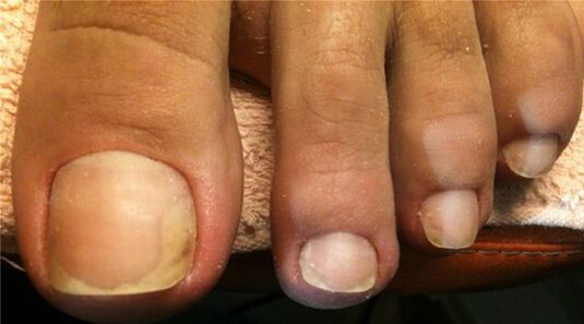
This form of disease is most common.In most cases, the cause of onychomycosis is dermatomycetes, especially the Trichophyton rubrum.The pathogens penetrate the nail plate from the side of the free wind and the side edges.Perenophaeum is hyperkeratosis, which results in the nail detached from the finger tissues (onycholysis), loses transparency, becomes whitish or yellow, and begins to crumble.As the submarine develops hyperkeratosis, the nail plate seems thickened.As the disease progresses, the focus of the lesion spreads to the hole, as indicated by the emerging yellow strips.Over time, the entire nail plate and the matrix are involved in the pathological process, which will over time lead to dystrophy and nail destruction.
In elderly people, it is often observed that the pronounced hyperkeratosis (thickening), onychogrifosis (thickening and deformation of poultry in the form of poultry claws), or koilonichia (concave deformation).Their nails are often affected by mixed flora - dermatophytes, shapes and even bacteria.

The surface (white) of onychomycosis on the legs
White surface onycommicosis on the legs is the second largest form of damage to the damage.This is mainly due to the Trichophyton mentagrophytes var.The interdigitale that penetrates directly into the nail plate on the upper (pre-r) and some types of fungal hectophytane.Mostly the nail on the first finger of the foot, less often - the fifth.
At first, small white spots and strips appear on their surface, which eventually captures the growing surface.Gradually the color will be yellow, ocher.The surface of the nail is relaxed, coarse, dusty, easily jumps.Compression and separation of nail beds does not occur.
The proximal submarine form of onychomycosis on the legs
This form of mycosis is a rarity.All onychomycosis approx.Make up 3%.This is due to Candida Albicans and Trichophyton Rubrum yeast -like mushrooms.Candidiasis of the nail was preceded by inflammation of the perologic rollers.It swells, gets red, becomes brilliant.The cuticle is lifted and the infection penetrates the last part of the matrix and the nail bed when the cracks of the nail plate and nail plate are damaged, the loss of natural glow and accumulation.Gradually the nail is destroyed and disappears in severe cases.This form of onychomycosis is often found in HIV-infected patients.
A complete distrophical form of onychomycosis on the legs
This form of onychomycosis is more common with a long -term (chronic course) disease, which is more common in Trichophyton rubrum and Candida albicans fungi.At the same time, the nail plate, bed and matrix are involved in the pathological process.Nail forgiveness occurs as a result of the developing hyperkeratosis of the submarine.Over time, the nail plate will be destroyed and the new matrix affected will not increase or grow badly.

Types of damage to nail plates
3 Opportunities for onychomycosis:
- Normotorophical.
- Hypertrophic.
- Atrophic.
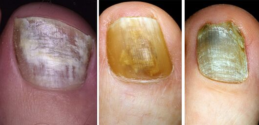
Normotrophic type onychomycosis on the legs
Normotrophic type of infection is localized in the upper layers of the nail plate.Its thickness and color do not change in the disease, but the stains and strips are visible in the depths.The color of the nails turns to yellow in white.After a while, the stains and strips merge.The injury area spreads to the entire nail plate except the moon.Fracture and crying are not observed.Occasionally, a slight relaxation of the free edge is detected.Proper treatment is possible to cure.
Hypertrophic type onychomycosis on the legs
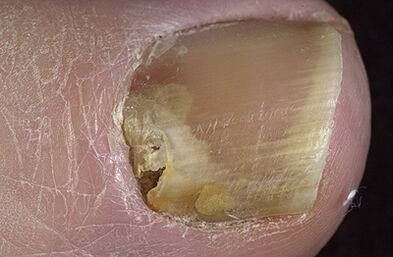
This type of onychomycosis is the most common.As a result of the development of the submarine hyperkeratosis, the nail plate is significantly thickened, deformed and loses its glow.The nails become uneven, boring, brownish-gray and crumbling.The area of the moon is not affected.The disease causes tangible discomfort to the patient.In elderly patients, the development of onychogrifosis is observed - the nails thicken, extend and bow like a bird's arm.
Atrophic type onychomycosis on the legs
The atrophic (onycholitic) type nail plate quickly loses its connection from the nail bed, many cavities appear in its layers, fades, becomes thinner, and turns the color to whitish or yellow and white.The surface remains smooth for a long time.Over time, partial destruction occurs.
Signs and symptoms of nail fungus
Most often, the nail change begins with a free (distal) or side (side) edge.
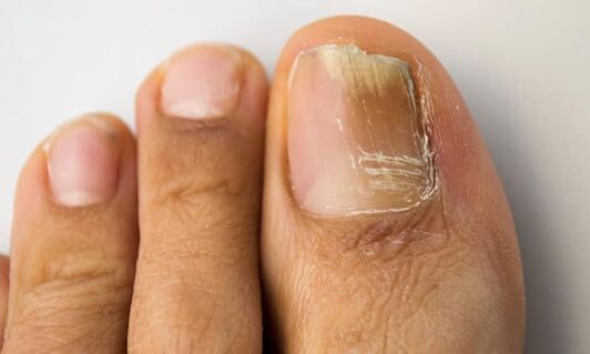
Color change.In the case of onychomycosis, a change in the color of the nail plate is the first sign of the disease.It becomes transparent, often loses its glow, white or yellow, overlapping mold - brown, brown, green and even black.
Compression.The increase in the number of horny masses results in the thickening of the nail as a result of the developing hyperkeratosis of the submarine.
Crushing and destruction.As a result of the vital activity of fungi, the nail plate first collapses and is completely destroyed over time.
Nail damage features different types of Onychomycosis
Damage to nails with different types of fungal diseases has its own properties.The main types of pathogens are Trichophyton Rubrum (70 - 90%) and Trichophyton mentagrophytes V.Intigitale (8 - 30%).Candida albicans, mold, T. Mentagrophytes V.gypseum, T. Verrucosum, T. Tonsuras and T. Visaceum, Epidermophyton Floccosum, Trichophyton is much less common.Schonleinii.
Onychomycosis on the legs Rubrophytia
The Russian Federation Rubrophytics accounts for 70-90% of all microsis.The leg of the disease are most often affected (usually a squamous dry type).The legs rubrofitia are an essential satellite of the feet nail fungus.In mycosis, the distal-dilutal form of onychomycosis is generally formed, the pronounced hyperkeratosis is typical, with several fingers on the foot at the same time and often fingers on the one hand.The disease occurs without specific subjective feelings.Pain and discomfort occurs with outstanding hyperkeratosis, onichogrifosis and increasing nails when wearing shoes.The source of infection is often found in the patient's family.
Often related onychomycosis are recorded: Trichophyton Rubrum and Candida Albicans, Trichophyton rubrum and molds.It is important to assess a cultural study.
Onychomycosis on the legs t.Mentagrophytes mushrooms.V.Intigitale
Mushroom T. Mentagrophytes.V.Intigitale affects the skin of the legs and nails.Epidermophytosis accounts for 10-30% of total micoses of the legs.
In the case of the disease, it affects the upper (back) part of the nail plate.The superficial white form of onychomycosis is usually formed.The pathological process is primarily involved in the 1 and 5 toes (while walking the shoes under the largest trauma) and 1 and 4 packaging folds.The spread of the infection occurs when a common bath, in the shower, in the sauna, in the pool, on the beaches and in the pools.
Onychomycosis on the legs with damage to the Candida genus yeast
This form of legs is a rarity.This accounts for less than 3% of all onychomycosis.The disease is often recorded in people with chronic generalized candidiasis.Nail damage usually begins with inflammation of the perologate roller near the hole.Edema and thickening can be observed, leading to the separation of the cuticle from the surface of the plate.In addition, fungi fall freely into the matrix and nail bed (proximal sub-nibble shape), when furrows, irregularities and cracks appear in the nail, the loss of natural glow and accumulation appears, a brownish brown color.Gradually the nail is destroyed and disappears in severe cases.
Onychomycosis on the legs caused by molds
Plastic mushrooms are placed in a nail already affected, in the spaces between the nail bed or the deserted vessels.The hyperkeratosis then develops and slowly destroyed the nail plate, which is painted in black (Scytalidium spp.) Or Green or gray (scopulariopsis brevicaulis) during the disease.
Diagnosis of onychomycosis
Diagnosis of onychomycosis is based on epidemiological history, clinical picture of the disease and the data of the laboratory research method.
Microscopic examination of the substance develops the nature of the disease (fungus or other pathogen).Fungi are identified by microbiological examination (plants of a substance on the medium), later microscopy of pure culture.The process is tiring, success is achieved in half of the cases.The key to a successful microbiological examination is the proper collection of materials from the affected nails.
Differential diagnosis
Only half of the cases of patients with changes in the nails and changes in color.Onychomycosis should be distinguished from eczema, psoriasis, reitera syndrome, pachionichia, daria, flat zuzmot, Norwegian cracks, bacterial lesions.